LL-37
LL-37 is the only human cathelicidin antimicrobial peptide, playing a critical role in innate immunity through direct antimicrobial activity, immunomodulation, wound healing, and anti-biofilm properties.
Overview
LL-37 derives its name from its 37-amino acid length and the two leucine residues at its N-terminus. It is encoded by the CAMP gene (cathelicidin antimicrobial peptide) and is the C-terminal fragment of hCAP18, released through proteolytic cleavage by proteinase 3 in neutrophils and by kallikreins in keratinocytes. The peptide adopts an amphipathic alpha-helical structure in physiological environments, which is essential for its membrane-disrupting antimicrobial mechanism.
Beyond direct antimicrobial killing, LL-37 functions as an immune alarm signal — it acts as a chemoattractant for neutrophils, monocytes, and T cells, modulates toll-like receptor (TLR) signaling, stimulates angiogenesis, and promotes wound re-epithelialization. Vitamin D is a primary transcriptional regulator of CAMP gene expression, linking vitamin D status directly to LL-37 production and innate immune competence.
Mechanism of Action
LL-37 exerts its biological effects through multiple mechanisms:
- Direct membrane disruption: The amphipathic alpha-helical structure allows LL-37 to insert into microbial membranes, forming pores or disrupting membrane integrity through a "carpet" model. This mechanism is effective against gram-positive and gram-negative bacteria, fungi, and enveloped viruses Dürr et al. (2006).
- Anti-biofilm activity: LL-37 inhibits biofilm formation at sub-inhibitory concentrations by downregulating genes involved in biofilm attachment and quorum sensing. It is effective against biofilms formed by Pseudomonas aeruginosa, Staphylococcus aureus, and Staphylococcus epidermidis Overhage et al. (2008).
- Immunomodulation: LL-37 activates the formyl peptide receptor-like 1 (FPRL1/FPR2), chemokine receptors, and P2X7 purinergic receptors on immune cells, triggering chemotaxis, cytokine modulation, and inflammasome activation. It suppresses LPS-induced pro-inflammatory cytokine release while enhancing anti-inflammatory responses Scott et al. (2002).
- Wound healing: LL-37 promotes keratinocyte migration, proliferation, and re-epithelialization through transactivation of the epidermal growth factor receptor (EGFR). It also stimulates angiogenesis via FPRL1 activation on endothelial cells Heilborn et al. (2003).
- Nucleic acid sensing: LL-37 binds to self-DNA and self-RNA, forming condensed complexes that are internalized by plasmacytoid dendritic cells to trigger TLR9- and TLR7-mediated type I interferon responses. This mechanism is implicated in the pathogenesis of psoriasis Lande et al. (2007).
Reconstitution Calculator
LL-37
**LL-37** is a 37-amino acid cationic antimicrobial peptide and the sole catheli
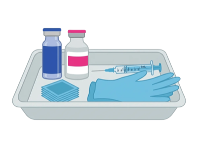
Set up a clean workspace with all supplies ready.
7x / week for weeks
This calculator is provided for informational and educational purposes only. It is not intended as medical advice, diagnosis, or treatment guidance. Always consult a qualified healthcare professional before preparing or administering any substance. PepGuide assumes no liability for decisions made based on these calculations.
Research
Vitamin D-Cathelicidin Axis and Tuberculosis
The landmark discovery linking vitamin D, cathelicidin, and antimicrobial immunity came from Liu et al. (2006), who showed that TLR2/1 activation on human macrophages induces VDR and CYP27B1, enabling local synthesis of 1,25(OH)₂D₃ from circulating 25(OH)D. This active vitamin D metabolite drives CAMP transcription and LL-37 production, which mediates intracellular killing of M. tuberculosis. Critically, this pathway requires sufficient circulating 25(OH)D — serum levels below 20 ng/mL result in inadequate LL-37 induction and impaired mycobacterial killing Liu et al. (2006). This finding provided the molecular explanation for the historical efficacy of sunlight and cod liver oil in tuberculosis treatment.
Skin Infections and Barrier Defense
Cathelicidin is a key component of skin antimicrobial defense. hCAP-18/LL-37 is constitutively expressed in keratinocytes and upregulated during infection, wounding, and UV exposure. Patients with atopic dermatitis have reduced cathelicidin expression, which contributes to their increased susceptibility to skin infections by S. aureus and eczema herpeticum Ong et al. (2002). Conversely, psoriatic skin overexpresses cathelicidin, but the LL-37 produced can form complexes with self-DNA that activate plasmacytoid dendritic cells, driving type I interferon production and autoimmune inflammation Lande et al. (2007).
Rosacea Connection
Yamasaki et al. (2007) demonstrated that rosacea skin contains abnormally high levels of cathelicidin and abnormally processed LL-37 fragments due to elevated KLK5 activity. These aberrant cathelicidin peptides are more potent inducers of inflammation and angiogenesis than native LL-37, directly contributing to the erythema, inflammation, and telangiectasia characteristic of rosacea. KLK5 inhibition reduces rosacea-like inflammation in mouse models, establishing cathelicidin processing as a therapeutic target Yamasaki et al. (2007).
Immunomodulation Beyond Antimicrobial Activity
The immunomodulatory functions of cathelicidin/LL-37 extend well beyond direct microbial killing. LL-37 neutralizes LPS and prevents endotoxin-induced septic shock. It modulates macrophage polarization, promotes autophagy, enhances neutrophil extracellular trap (NET) formation, and regulates apoptosis of immune cells. These pleiotropic effects position cathelicidin as a central mediator of the innate immune response rather than simply an antibiotic peptide Zanetti (2004).
Anti-Tumor Role in Colon Cancer
LL-37 expression is significantly reduced in colon cancer tissues compared to adjacent normal mucosa. Ren et al. (2012) demonstrated that exogenous LL-37 inhibits colon cancer cell proliferation (HCT116, HT-29, LoVo cell lines) in a dose-dependent manner, induces caspase-dependent apoptosis, and suppresses colony formation. The anti-tumor mechanism involves both direct membrane-mediated cytotoxicity and activation of intrinsic apoptotic pathways. In vivo, intratumoral injection of LL-37 reduced colon cancer xenograft growth in nude mice. The downregulation of LL-37 in colon cancer is attributed to epigenetic silencing of the CAMP promoter through DNA methylation Ren et al. (2012). These findings support the hypothesis that LL-37 functions as a tumor suppressor in the colonic epithelium and provide a mechanistic link between vitamin D's protective effect against colon cancer and cathelicidin expression.
Anti-Tumor Role in Gastric Cancer
Li et al. (2013) reported that LL-37 expression is downregulated in human gastric cancer specimens and that exogenous LL-37 inhibits gastric cancer cell proliferation, induces mitochondrial-mediated apoptosis, and suppresses tumor growth in xenograft models. The anti-tumor mechanism in gastric cancer is mediated through p53-dependent pathways, with LL-37 stabilizing p53 protein and promoting its nuclear translocation. Additionally, LL-37 inhibits proteasome activity in gastric cancer cells, leading to accumulation of pro-apoptotic proteins Li et al. (2013).
Wound Healing
LL-37 is highly expressed at wound margins and is critical for normal cutaneous repair. Heilborn et al. showed that chronic non-healing venous leg ulcers are deficient in LL-37 compared to normally healing acute wounds, implicating cathelicidin deficiency in impaired wound repair. LL-37 promotes healing through antimicrobial protection of the wound bed, keratinocyte migration via EGFR transactivation, and angiogenesis stimulation Heilborn et al. (2003).
Pro-Tumor Role in Ovarian Cancer
In stark contrast to colon cancer, LL-37 is significantly overexpressed in ovarian tumor tissues, with both cancer cells and tumor-infiltrating macrophages serving as sources. Coffelt et al. (2008, 2009) established that LL-37 functions as a growth factor in ovarian cancer, promoting proliferation through EGFR transactivation, enhancing migration and invasion through FPR2-MEK/ERK signaling, and increasing chemoresistance to cisplatin and paclitaxel. Critically, LL-37 recruits mesenchymal stem cells to the tumor microenvironment, which differentiate into tumor-associated fibroblasts that further support tumor growth through paracrine signaling Coffelt et al. (2009). Neutralizing antibodies against LL-37 inhibited ovarian tumor growth in mouse models, confirming the peptide's functional role in promoting tumor progression Coffelt et al. (2008).
Angiogenesis and Vascularization
Koczulla et al. (2003) identified LL-37 as a pro-angiogenic factor, demonstrating that the peptide promotes endothelial cell proliferation, migration, and neovascularization both in vitro and in vivo (chick chorioallantoic membrane and rabbit hindlimb ischemia models). The angiogenic effect is mediated through FPR2 on endothelial cells and involves activation of the MAPK/ERK pathway. In the context of cancer, this pro-angiogenic activity provides a mechanism by which LL-37-overexpressing tumors can promote their own vascularization, supporting nutrient delivery and metastatic spread Koczulla et al. (2003).
Immunomodulatory Effects in the Tumor Microenvironment
LL-37's immunomodulatory effects add another layer of complexity to its role in cancer. The peptide can recruit and activate multiple immune cell types: neutrophils and monocytes (via FPR2), immature dendritic cells (via FPR2 and CCR6-like mechanisms), and T cells. In some tumor contexts, LL-37 promotes anti-tumor immunity by enhancing NK cell cytotoxicity and dendritic cell maturation. In others, LL-37 promotes immunosuppression by driving macrophage polarization toward the M2 (tumor-associated) phenotype and expanding regulatory T cells. The net immunological effect depends on the local cytokine milieu, tumor type, and LL-37 concentration Kuroda et al. (2015).
Ongoing & Future Research
- OP-145 (P60.4Ac): Truncated LL-37 analog in Phase 2 clinical trials for chronic otitis media (NCT02022540)
- Development of D-amino acid LL-37 analogs resistant to protease degradation with improved therapeutic index
- LL-37-functionalized wound dressings and biomaterial coatings for medical device anti-infective applications
- Research into LL-37-based peptides for antibiotic-resistant infections (MRSA, carbapenem-resistant Enterobacteriaceae)
- Investigation of LL-37 analogs with reduced hemolytic activity and improved selectivity index
- LL-37 as a cancer therapeutic: ongoing research into analogs that retain anti-tumor activity without pro-tumorigenic EGFR effects
- Vitamin D supplementation trials using LL-37 levels as a biomarker for innate immune competence
- LL-37-derived peptides for anti-biofilm coatings on catheters and prosthetic implants (Zhang et al., DOI: 10.1016/j.peptides.2024.171156)
Antimicrobial Activity
LL-37 demonstrates broad-spectrum antimicrobial efficacy. It kills both gram-positive organisms (S. aureus, S. pyogenes) and gram-negative pathogens (E. coli, P. aeruginosa, K. pneumoniae) at micromolar concentrations. It is also effective against mycobacteria, including Mycobacterium tuberculosis, where vitamin D-induced LL-37 expression is a key mechanism of macrophage antimicrobial defense Liu et al. (2006). Antifungal activity against Candida albicans and antiviral effects against influenza, HIV, and herpes simplex virus have also been documented Barlow et al. (2011).
Anti-Biofilm Properties
Bacterial biofilms are a major clinical challenge due to their resistance to conventional antibiotics. LL-37 has shown the ability to prevent and disrupt biofilm formation by P. aeruginosa at concentrations well below its minimum inhibitory concentration (MIC) for planktonic bacteria. Overhage et al. demonstrated that LL-37 at 0.5 μg/mL inhibited P. aeruginosa biofilm formation by 40%, reduced pre-formed biofilm thickness, and stimulated twitching motility that disrupted biofilm architecture Overhage et al. (2008).
Wound Healing and Skin
LL-37 is upregulated at wound sites and plays a critical role in cutaneous repair. It is highly expressed in the wound edge epithelium and promotes keratinocyte migration through EGFR transactivation. Heilborn et al. showed that chronic non-healing ulcers are deficient in LL-37 compared to acute healing wounds, suggesting that LL-37 deficiency contributes to impaired wound healing Heilborn et al. (2003). In skin conditions, LL-37 overexpression is associated with rosacea, where aberrant processing of hCAP18 by kallikrein 5 generates pro-inflammatory LL-37 fragments Yamasaki et al. (2007).
Cancer Research
LL-37 has demonstrated context-dependent effects in cancer biology. It exhibits direct cytotoxic effects against certain cancer cell lines, including gastric cancer and hematological malignancies, through membrane disruption and apoptosis induction. However, in some cancer types (ovarian, breast, lung), LL-37 may promote tumor growth through EGFR activation and angiogenesis stimulation. This duality underscores the complexity of LL-37's role in oncology and the need for cancer type-specific investigation Wu et al. (2010).
Vitamin D Connection
The link between vitamin D and innate immunity is mediated largely through LL-37. Vitamin D receptor (VDR) activation directly upregulates CAMP gene transcription. Liu et al. demonstrated that TLR2 activation in macrophages upregulates VDR and CYP27B1 (the enzyme converting 25(OH)D to active 1,25(OH)₂D), leading to LL-37 induction and intracellular killing of M. tuberculosis. This landmark study established the molecular basis for the historical use of sunlight and cod liver oil in tuberculosis treatment Liu et al. (2006).
Pro-Tumor Role in Breast and Lung Cancer
LL-37 promotes breast cancer progression through EGFR transactivation and downstream activation of the HER2/ErbB2 signaling network. Weber et al. (2009) showed that LL-37 stimulates breast cancer cell migration and invasion through metalloproteinase-dependent release of EGFR ligands, and that LL-37 expression is elevated in breast tumor tissues Weber et al. (2009). In non-small cell lung cancer (NSCLC), von Haussen et al. (2008) demonstrated that LL-37 is overexpressed in tumor tissues and acts as an autocrine/paracrine growth factor, promoting NSCLC cell proliferation through FPRL1/FPR2 signaling and EGFR transactivation. LL-37 also enhances lung cancer cell migration and invasion, suggesting a role in metastatic progression von Haussen et al. (2008).
Neurological/Immunological Mechanisms
Membrane disruption mechanisms:
- Carpet model: LL-37 monomers align parallel to the membrane surface, covering it like a carpet until a critical concentration is reached, causing membrane disintegration
- Toroidal pore model: At higher concentrations, LL-37 inserts perpendicular to the membrane, forming transmembrane pores with lipid headgroups lining the pore interior
- The amphipathic alpha-helix is essential: hydrophobic face interacts with lipid acyl chains; hydrophilic/cationic face interacts with anionic lipid headgroups
- Selectivity for microbial membranes: bacterial membranes are enriched in anionic phospholipids (phosphatidylglycerol, cardiolipin) while mammalian membranes are enriched in zwitterionic phospholipids (phosphatidylcholine) and cholesterol
Immunomodulatory receptor signaling:
- FPRL1/FPR2 (formyl peptide receptor-like 1): Primary receptor for LL-37's immunomodulatory effects. Activation → Gi-coupled signaling → chemotaxis of neutrophils, monocytes, and T cells to infection sites (Scott et al., PMID: 11796220)
- P2X7 purinergic receptor: LL-37 activates P2X7 → IL-1β processing and release via NLRP3 inflammasome activation. This links LL-37 to inflammasome-mediated innate immune responses.
- EGFR transactivation: LL-37 → metalloproteinase activation → HB-EGF release → EGFR phosphorylation → keratinocyte migration and proliferation (wound healing) (Heilborn et al., PMID: 12932393)
- TLR9 (indirect): LL-37-DNA complexes activate endosomal TLR9 in plasmacytoid DCs → type I IFN production. This is a key pathogenic mechanism in psoriasis but also contributes to antiviral defense (Lande et al., PMID: 17676033)
Anti-biofilm mechanisms:
- At sub-MIC concentrations: downregulates biofilm-specific genes (lasI, lasR quorum sensing regulators in P. aeruginosa)
- Stimulates type IV pilus-dependent twitching motility → disrupts biofilm architecture (Overhage et al., PMID: 18227163)
- Prevents initial bacterial adhesion to surfaces
- Disrupts pre-formed biofilms through charge-mediated interactions with the biofilm exopolysaccharide matrix
Vitamin D-LL-37 axis (molecular detail):
- TLR2/1 activation on macrophages → upregulates VDR and CYP27B1
- CYP27B1 converts 25(OH)D → active 1,25(OH)₂D locally
- 1,25(OH)₂D binds VDR → VDR/RXR heterodimer binds VDRE (vitamin D response element) in CAMP gene promoter
- CAMP transcription → hCAP18 protein → proteolytic processing → mature LL-37 peptide
- This pathway requires sufficient circulating 25(OH)D substrate — explaining why vitamin D deficiency impairs antimicrobial immunity (Liu et al., PMID: 16497887)
Safety Profile
LL-37 research has identified several safety considerations:
- Hemolytic activity: At higher concentrations (>25 μM), LL-37 can exhibit hemolytic effects on human erythrocytes due to its membrane-active mechanism. Therapeutic concentrations are typically well below this threshold.
- Pro-inflammatory potential: In the context of autoimmune conditions such as psoriasis, LL-37 can amplify inflammation through the LL-37-DNA/RNA complex-mediated activation of plasmacytoid dendritic cells and type I interferon production.
- Rosacea association: Aberrant LL-37 processing in the skin is implicated in the pathogenesis of rosacea, suggesting caution in dermatological applications.
- Cytotoxicity: Dose-dependent cytotoxicity to host cells has been observed at concentrations exceeding those required for antimicrobial activity.
- Serum inactivation: LL-37 activity is partially inhibited by serum proteins and high salt concentrations, which may limit efficacy in certain physiological environments.
Research into truncated LL-37 analogs and engineered derivatives aims to improve the therapeutic index by reducing host cell toxicity while retaining antimicrobial and immunomodulatory properties.
Pharmacokinetic Profile
- Half-life
- Short (rapid proteolytic degradation)
- Tmax
- 30 min
- Protein Binding
- LL-37 binds to serum proteins (apolipoprotein A-I, albumin), which partially protects it from degradation but also reduces free active concentration. Serum significantly reduces antimicrobial activity compared to buffer conditions.
- Distribution
- Produced locally by neutrophils (stored in specific granules as hCAP18), macrophages, epithelial cells, and keratinocytes. Released at sites of infection, inflammation, and wounding.
Quick Start
- Typical Dose
- 0.5-1.6 mg/mL topical gel or 100-150 mcg injectable
- Frequency
- Topical: Daily to twice weekly on wounds. Injectable: Once daily subcutaneous
- Route
- Topical, Subcutaneous injection, Intravenous
- Cycle Length
- 2-8 weeks depending on wound healing progress
- Storage
- Lyophilized: 2-8°C. Reconstituted: 2-8°C, maintain sterility
Molecular Structure
- Formula
- C₂₀₅H₃₄₀N₆₀O₅₃
- Weight
- 4493.3 Da
- Length
- 37 amino acids
- CAS
- 154947-66-7
- PubChem CID
- 16198951
- Exact Mass
- 4492.5821 Da
- LogP
- -20.7
- TPSA
- 1900 Ų
- H-Bond Donors
- 68
- H-Bond Acceptors
- 65
- Rotatable Bonds
- 166
- Complexity
- 10700
Identifiers (SMILES, InChI)
InChI=1S/C205H340N60O53/c1-20-114(16)162(261-179(296)128(66-40-46-86-211)235-176(293)135(74-78-155(273)274)243-170(287)125(63-37-43-83-208)241-192(309)148(106-266)257-174(291)126(64-38-44-84-209)234-171(288)129(67-47-87-225-201(215)216)240-185(302)142(98-119-55-29-24-30-56-119)252-187(304)144(100-121-59-33-26-34-60-121)253-189(306)146(102-158(279)280)233-154(272)104-230-167(284)138(94-109(6)7)248-166(283)122(212)93-108(4)5)194(311)231-105-153(271)232-123(61-35-41-81-206)168(285)242-136(75-79-156(275)276)177(294)251-141(97-118-53-27-23-28-54-118)184(301)238-124(62-36-42-82-207)169(286)236-131(69-49-89-227-203(219)220)181(298)263-164(116(18)22-3)197(314)259-160(112(12)13)195(312)246-134(73-77-151(213)269)175(292)237-132(70-50-90-228-204(221)222)180(297)262-163(115(17)21-2)196(313)245-127(65-39-45-85-210)172(289)256-147(103-159(281)282)190(307)254-143(99-120-57-31-25-32-58-120)186(303)249-139(95-110(8)9)183(300)239-130(68-48-88-226-202(217)218)173(290)255-145(101-152(214)270)188(305)250-140(96-111(10)11)191(308)260-161(113(14)15)199(316)265-92-52-72-150(265)193(310)244-133(71-51-91-229-205(223)224)182(299)264-165(117(19)268)198(315)247-137(76-80-157(277)278)178(295)258-149(107-267)200(317)318/h23-34,53-60,108-117,122-150,160-165,266-268H,20-22,35-52,61-107,206-212H2,1-19H3,(H2,213,269)(H2,214,270)(H,230,284)(H,231,311)(H,232,271)(H,233,272)(H,234,288)(H,235,293)(H,236,286)(H,237,292)(H,238,301)(H,239,300)(H,240,302)(H,241,309)(H,242,285)(H,243,287)(H,244,310)(H,245,313)(H,246,312)(H,247,315)(H,248,283)(H,249,303)(H,250,305)(H,251,294)(H,252,304)(H,253,306)(H,254,307)(H,255,290)(H,256,289)(H,257,291)(H,258,295)(H,259,314)(H,260,308)(H,261,296)(H,262,297)(H,263,298)(H,264,299)(H,273,274)(H,275,276)(H,277,278)(H,279,280)(H,281,282)(H,317,318)(H4,215,216,225)(H4,217,218,226)(H4,219,220,227)(H4,221,222,228)(H4,223,224,229)/t114-,115-,116-,117+,122-,123-,124-,125-,126-,127-,128-,129-,130-,131-,132-,133-,134-,135-,136-,137-,138-,139-,140-,141-,142-,143-,144-,145-,146-,147-,148-,149-,150-,160-,161-,162-,163-,164-,165-/m0/s1
POIUWJQBRNEFGX-XAMSXPGMSA-NResearch Indications
Wound Healing
68% ulcer area reduction demonstrated with 0.5mg/mL concentration in clinical trials.
Enhanced granulation tissue formation and wound closure.
Accelerated healing demonstrated in animal models.
Immunity
Boosts natural immune defenses against pathogens.
Active against MRSA and multi-drug resistant bacteria with >4 log reduction in biofilms.
Balances immune responses without causing immunosuppression.
Skin Health
Stimulates skin cell growth and regeneration.
Promotes new blood vessel formation for tissue repair.
Supports collagen production for wound healing.
Research Protocols
topical
Topical application is the most established route, providing direct access to wound sites with minimal systemic exposure.
| Goal | Dose | Frequency | Duration |
|---|---|---|---|
| Wound healing - Standard | 0.5mg/mL gel/cream | Daily to twice weekly | —(Route: Direct wound application) |
| Wound healing - Intensive | 1.6mg/mL gel/cream | Daily | —(Route: Direct wound application) |
Reconstitution Guide (mg vial + mL BAC water)
- Clean and debride wound area
- Apply thin layer directly to wound surface
- Cover with sterile dressing if needed
- Change per clinical protocol (daily to twice weekly)
- Monitor healing progress and adjust frequency
- Document wound measurements and progression
subcutaneous Injection
Antimicrobial peptide with wound-healing properties. Slow weekly titration from 50 mcg to 400 mcg.
| Goal | Dose | Frequency | Duration |
|---|---|---|---|
| Week 1 | 50 mcg | Once daily | Week 1 |
| Week 2 | 100 mcg | Once daily | Week 2 |
| Week 3-4 | 150-200 mcg | Once daily | Weeks 3-4 |
| Full dose | 300-400 mcg | Once daily | Weeks 5-8+(Titrate ~50 mcg/week. Optional 5-on/2-off schedule. Cycle: 8-12 weeks.) |
Reconstitution Guide (5mg vial + 3mL BAC water)
- Wipe vial tops with alcohol swab
- Draw 3.0 mL bacteriostatic water into syringe
- Inject slowly down the inside wall of the peptide vial
- Gently swirl to dissolve — never shake
- Resulting concentration: 1.67 mg/mL
- For 100 mcg dose: draw 6 units (0.06 mL)
- For 200 mcg dose: draw 12 units (0.12 mL)
- For 400 mcg dose: draw 24 units (0.24 mL)
- Store reconstituted vial refrigerated at 2-8°C
Interactions
Peptide Interactions
Enhanced wound healing through complementary mechanisms.
Combined anti-inflammatory and healing effects.
Acts as cofactor for endogenous LL-37 production.
Both promote wound healing through different pathways — LL-37 via EGFR transactivation and antimicrobial defense; GHK-Cu via matrix metalloproteinase modulation and collagen synthesis.
Cathelicidin (LL-37) and beta-defensins are the two major families of human antimicrobial peptides, both induced by vitamin D signaling via VDR-bound vitamin D response elements. They are co-expressed in keratinocytes and immune cells, providing complementary antimicrobial defense — LL-37 through membrane pore formation and defensins through electrostatic disruption. Both also function as immune signaling molecules recruiting leukocytes and stimulating cytokine production (Gombart, 2009).
Alpha-defensins (HNP1-4) from neutrophils and cathelicidin LL-37 represent complementary innate immune effectors. Both act as chemoattractants for dendritic cells, T cells, monocytes, and neutrophils. They are co-released from neutrophil granules during infection. Alpha-defensins provide antimicrobial activity in the phagosome while LL-37 operates at mucosal surfaces. Together they bridge innate and adaptive immunity (Yang et al., 2002).
Safe combined use with no negative interactions.
Complementary innate (LL-37) + adaptive (Ta1) immune enhancement. LL-37 provides direct antimicrobial killing and immune cell chemotaxis; Ta1 enhances T-cell and NK cell function. Theoretical synergy for immunocompromised patients.
Both LL-37 and alpha-MSH are antimicrobial peptides co-expressed in inflamed skin (e.g., hidradenitis suppurativa). LL-37 is released upon NF-kB-induced inflammatory signals and activates innate immune recruitment, while alpha-MSH suppresses NF-kB and downregulates pro-inflammatory cytokines. They provide balanced immune regulation: LL-37 for pathogen clearance and alpha-MSH for inflammation resolution. No antagonistic interaction; complementary defense mechanisms (Singh & Singh, 2014).
KPV (alpha-MSH C-terminal tripeptide) suppresses NF-kB activation and pro-inflammatory cytokines, while cathelicidin LL-37 is induced by NF-kB inflammatory signaling for antimicrobial defense. These represent complementary aspects of host defense — KPV limits excessive inflammation while LL-37 provides direct antimicrobial killing. No pharmacological antagonism; both contribute to mucosal and skin immunity through distinct melanocortin and innate immune pathways.
Interaction profile not established; limited data available for most combinations.
Substance P triggers mast cell degranulation and neurogenic inflammation, which can upregulate cathelicidin expression in keratinocytes. In inflammatory skin conditions like rosacea, excessive cathelicidin processing by kallikrein-5 produces pro-inflammatory LL-37 fragments. SP-driven neurogenic inflammation may amplify this pathological cathelicidin processing. Monitor for exacerbation of inflammatory skin conditions when both pathways are active.
What to Expect
What to Expect
Reduced bacterial load, initial wound bed preparation
Increased granulation tissue formation, epithelial migration
Significant wound size reduction
Continued healing toward wound closure
Safety Profile
Common Side Effects
- Generally well-tolerated
- Mild injection site reactions (injectable)
- Local irritation at application site (topical)
Contraindications
- Pre-existing hypersensitivity to peptides
- Compromised immune status (relative contraindication)
- Non-sterile wound environments
Discontinue If
- Severe injection site reactions with spreading inflammation
- Allergic reaction signs (rash, difficulty breathing, swelling)
- Persistent flu-like symptoms
- Signs of wound infection (increased redness, warmth, drainage)
- Worsening wound condition or delayed healing
- Unusual pain or irritation at application site
Quality Indicators
What to look for
- Sterile, clear formulation
- ≥98% purity by HPLC
- White to off-white lyophilized powder
- Proper cold chain maintenance (2-8°C)
- Appropriate gel base compatible with peptide activity
- Clinical-grade concentrations (0.5-1.6mg/mL)
Caution
- Brief room temperature exposure acceptable if promptly refrigerated
- Homemade topical formulations may lack sterility
Red flags
- Discolored or cloudy solutions
- Yellow discoloration or visible particles
- Pre-mixed liquid formulations (LL-37 unstable long-term)
- Non-sterile preparations
- Microbial contamination
Frequently Asked Questions
References (27)
- [13]
- [1]Venous Leg Ulcer Trial - Grönberg et al. (2014)
- [2]Melanoma Phase I Trial (2015)
- [3]MRSA Biofilm Study - Noore et al. (2019)
- [4]Structural Analysis - Wang et al. (2008)
- [8]
- [12]
- [24]Coorens M et al — LL-37 in respiratory infections: antimicrobial mechanisms and immunomodulatory potential (2023)
- [25]Ridyard KE, Bhatt A, Bhatt N — Cathelicidin-derived antimicrobial peptides as next-generation antibiotics: challenges and opportunities (2023)
- [27]
- [28]
- [9]
- [10]
- [5]
- [11]
- [7]
- [20]Liu PT, Stenger S, Li H, et al Toll-like receptor triggering of a vitamin D-mediated human antimicrobial response Science (2006)
- [14]
- [15]Dürr UH, Sudheendra US, Ramamoorthy A LL-37, the only human member of the cathelicidin family of antimicrobial peptides Biochim Biophys Acta (2006)
- [16]Scott MG, Davidson DJ, Gold MR, et al The human antimicrobial peptide LL-37 is a multifunctional modulator of innate immune responses J Immunol (2002)
- [17]Overhage J, Campisano A, Bains M, et al Human host defense peptide LL-37 prevents bacterial biofilm formation Infect Immun (2008)
- [18]Heilborn JD, Nilsson MF, Kratz G, et al The cathelicidin anti-microbial peptide LL-37 is involved in re-epithelialization of human skin wounds J Invest Dermatol (2003)
- [19]Lande R, Gregorio J, Facchinetti V, et al Plasmacytoid dendritic cells sense self-DNA coupled with antimicrobial peptide Nature (2007)
- [21]Barlow PG, Svoboda P, Mackellar A, et al Antiviral activity and increased host defense against influenza infection elicited by the human cathelicidin LL-37 PLoS One (2011)
- [22]Yamasaki K, Di Nardo A, Bardan A, et al Increased serine protease activity and cathelicidin promotes skin inflammation in rosacea Nat Med (2007)
- [23]Wu WK, Wang G, Coffelt SB, et al Emerging roles of the host defense peptide LL-37 in human cancer and its potential therapeutic applications Int J Cancer (2010)
- [26]Minns D et al — The neutrophil antimicrobial peptide cathelicidin/LL-37 shapes the inflammatory response in human skin (2022)
LKEK (Vascular Bioregulator)
**LKEK** (Leu-Lys-Glu-Lys) is a synthetic tetrapeptide belonging to the Khavinson series of bioregulatory peptides developed at the Saint Petersburg Institute o
L-Serine
L-Serine is a non-essential amino acid that plays a crucial role in protein synthesis, neurotransmitter production, and sphingolipid formation in the central ne